MRI in Multiple Sclerosis Diagnosis: The Complete Patient Guide
🌟 Introduction: Why MRI Matters in MS Diagnosis
MRI (Magnetic Resonance Imaging) has changed the way that we diagnose and treat multiple sclerosis (MS). It is a safe, non-invasive method that takes pictures of the brain and spinal cord - allowing the identification of classic MS lesions. The sooner the lesion becomes visible, the better the care we can provide, and MRI allows us to view what is going on inside a person before symptoms make an uproar. 📸🧩
🧬 Understanding Multiple Sclerosis and Its Impact on the Nervous System
What is Multiple Sclerosis (MS)?
Multiple Sclerosis is an autoimmune disease which is chronic in nature will be wreaking havoc on the body's immune system by mistake; the immune system picks on its victim, the central nervous system which is the brain and spinal cord. 😟 The body considers your brain and spinal cord a threat and go into protect and attack mode on the central nervous system, specifically on the fatty coat called myelin, which is a protective fatty covering that insulates the nerve fibers, just like plastic on wires. Myelin when broken down, doesn't allow for messages from brain to body, causing all types of symptoms: fatigue, blurry vision, numbness, etc.
The Role of Myelin and Nerve Fiber Damage in MS
Myelin facilitates fast, smooth communication between your brain and the rest of your body. 🧠➡️💪 In MS, the immune system attacks this protective layer. This is called demyelination, and it leaves the nerve underneath exposed and at risk of damage. Eventually, this causes problems with movement, thinking, and mood. The more we understand this process, the better we can address MS - and that is where MRI is important!
🌀 MRI Fundamentals Explained for Patients
How MRI Technology Works
MRI is like a superhero tool for doctor’s. It gives them great pictures of your insides without any surgery. 🛡️ It uses very powerful magnets and radio waves (no radiation!). The MRI machine aligns the hydrogen atoms in your body to produce pictures with the pulses. When the atoms snap back, they give off signals. The signals are used to make detailed pictures of your tissues. MRI works especially well with soft tissues, like the brain and spinal cord - which is great for an MS diagnosis. 🧲🧠
MRI vs. CT and X-rays: What Makes MRI Special?
Quick recap:
- X-rays = great for bones 🦴
- CT scans = 3D view using radiation 🔍
- MRI = best for brain, spinal cord, muscles, and soft tissues 🌟
MRI doesn’t involve radiation, which is a big win for safety. Plus, it captures soft tissue detail like no other tool — making it the go-to choice for spotting MS-related nerve damage.
Safe and Radiation-Free ✨
One of MRI’s biggest perks? It’s radiation-free. That makes it ideal for:
- Kids 👶
- Pregnant people 🤰
- Anyone needing multiple scans 🌀
- Naturally, there are some things to consider before hand. If you have any metal implants, pacemakers, or don't like enclosed spaces, be sure to communicate this to your doctor. There are ways to work around this (open MRIs or use of sedation) and keep it safe and comfortable. 😊
🧩 The Role of MRI in Diagnosing MS

Why MRI is the Gold Standard in MS Diagnosis
When suspecting MS the first tool that will be used is an MRI- magnetic resonance imaging. MRI is the best equipment we have to detect any signs of MS-related changes in either the brain or spinal cord. 🧠🔍 MRI can detect lesions (damaged areas), even if the patient does not show any symptoms. This will help with detecting changes early and starting treatment as soon as possible.
What an MRI Can Reveal About MS
MRI can detect:
- Lesions in the brain/spinal cord caused by demyelination 🧠
- Active inflammation that may not yet be causing symptoms 🔥
- Disease progression over time
Each scan tells a story about how MS is affecting your nervous system.
Demyelination: How It Appears on MRI Scans
In demyelination, the damaged areas appear to have water accumulation in them, which can manifest as white areas in some scans, and dark areas in others, depending on the type of MRI. These patterns allow radiologists and neurologists to determine whether what they are observing is consistent with MS.
🔬 MRI Sequences Used in MS Evaluation

T1-Weighted Scans (With and Without Gadolinium)
- Without contrast: Shows darker (hypointense) spots — areas of permanent damage 😓
- With contrast: Highlights bright (enhancing) spots — showing where new inflammation is happening 🔥
T2-Weighted Scans
These scans show total lesion burden — old and new lesions combined. It helps visualize the overall effect MS has had over time. 🗺️
FLAIR (Fluid Attenuated Inversion Recovery) Imaging
FLAIR scans filter out background “noise” from cerebrospinal fluid, making it easier to see MS lesions in the brain’s white matter. 🎨
💉 Gadolinium-Based Contrast Agents (GBCAs) in MS MRI
Purpose of Gadolinium in Detecting Active Inflammation
Gadolinium is a special dye injected during some MRI scans. It helps highlight active inflammation — showing areas where the blood-brain barrier has been compromised by MS. 🌈
Benefits vs. Risks of GBCAs
GBCAs are very effective, however they come with potential risks, especially for people who have kidney issues or who are pregnant. Some gadolinium may remain in your body for months or years; the long-term effects are still being studied. ⚖️
FDA Guidance and Safer Options for Gadolinium Use
The FDA recommends using macrocyclic GBCAs like Dotarem, Gadavist, or ProHance — these are less likely to stick around in your body. Always ask your doctor if contrast is truly necessary for your scan. ✅ [Ref. 1]
🔍 Special MRI Use Cases in MS
MRI After a Clinically Isolated Syndrome (CIS)
If you’ve had just one MS-like episode, called CIS, MRI can help determine your risk of developing full-blown MS. More lesions = higher risk. 📈
Predicting Disease Risk from Initial MRI Findings
Your very first MRI can provide clues about how likely you are to have future relapses. Doctors can spot hidden lesions you didn’t know you had! 🕵️
Tracking Subclinical MS Progression Without New Symptoms
Sometimes MS progresses quietly. MRI helps catch it early, before symptoms become more serious. Early intervention = better outcomes! 🌟
🧭 Monitoring Disease Activity Post-Diagnosis
![]()
Using MRI to Guide Treatment Plans
Doctors use MRI scans to see how well your treatment is working — or if it needs a change. It's a great way to make informed decisions. 💊📊
How Often Should You Get an MRI with MS?
Depends! Some people need yearly scans; others less often. Your neurologist will tailor a schedule based on your symptoms, history, and meds. 🗓️
Comparing MRI Results Over Time
Having a baseline and comparing new scans to it is key. That’s why it’s ideal to use the same machine each time, if possible. 🖥️🧾 [Ref. 2]
🧲 MRI Machine Strength: Does It Matter?
Magnet Strength: 1.5T vs. 3T vs. Open MRIs
- 1.5 Tesla (T): Most common, widely available
- 3T: Higher detail, great for spotting subtle lesions 🔬
- Open MRI: More comfortable, but less detailed — use only when needed 😊
Pros and Cons of Open MRI for Claustrophobic Patients
Open MRIs are great if you’re anxious in tight spaces. Just know the image quality might not be as high. Still, comfort is key! 😌
Consistency in Scanning Machines for Long-Term Tracking
Sticking with one MRI machine helps your doctor accurately track disease progression. It’s like comparing apples to apples. 🍎🍎
📅 Preparing for Your MS MRI Scan
How to Mentally and Physically Prepare
- Wear comfy clothes 👕
- No metal (zippers, jewelry, etc.)
- Eat lightly before your appointment 🥗
- Practice deep breathing to stay calm 😌
Questions to Ask Your Doctor Before the Scan
- Do I need gadolinium contrast this time?
- How often should I be scanned?
- What kind of MRI machine will be used?
Understanding MRI Reports with Your Neurologist
Ask your doctor to walk you through the report. It’s okay if you don’t understand it all — that’s what they’re there for. Knowledge is power! 💪
🚀 Advanced MRI Technologies in MS Research
High-Field and Ultra-High-Field MRI in Clinical Trials
Research centers use super-powerful MRI machines (like 7T) to study MS in high detail. These are mostly for research, but they’re teaching us a lot. 🧪 [Ref. 3]
Diffusion Tensor Imaging (DTI) and MS
DTI is a special scan that maps nerve fiber pathways — useful for understanding MS damage in more depth. 🌐
Functional MRI (fMRI) and Cognitive Impact Studies
fMRI helps us understand how MS affects thinking and memory by tracking real-time brain activity. 🧠💭
🧾 Interpreting MRI Results and Lesion Load

What Are Lesions and Why Do They Matter?
Lesions = signs of nerve damage. More lesions, or lesions in important areas like the spinal cord, might mean more serious symptoms. 😔
Lesion Location and Symptom Correlation
Lesions in the optic nerve might cause vision loss. In the spinal cord, they can affect movement. Lesion location matters as much as the number. 🎯
“Silent” Lesions and Brain Plasticity
Some lesions never cause noticeable symptoms because your brain rewires itself to “work around” the damage. That’s called neuroplasticity. Amazing, right? 🤯
🧪 MRI and Other Diagnostic Tools: A Combined Approach
Evoked Potentials
These tests measure how your brain responds to stimuli — delays may point to MS-related damage. ⚡
Lumbar Puncture
Also called a spinal tap, this test checks your spinal fluid for MS markers like oligoclonal bands. 🧬
Comprehensive Neurological Exams
A good old-fashioned check-up can reveal a lot: balance, vision, reflexes, strength — all are key for diagnosis. 🩺
🔄 Common Misunderstandings About MS MRIs
Myth: Lesions Always Equal Symptoms
Not always. Some lesions don’t cause symptoms thanks to brain adaptation. 📉
Myth: Normal MRI Means No MS
You can still have MS even with a normal scan, especially in early stages. Other tests help fill in the gaps. 🔍
Clarifying Radiologist vs. Neurologist Roles
Radiologists read the scans. Neurologists connect that info to your health and symptoms. Teamwork makes the diagnosis work! 🤝
💪 Patient Advocacy: Taking Charge of Your Diagnostic Journey
Empowering Questions to Ask
- How many lesions do I have?
- Where are they located?
- Has anything changed since my last scan?
Finding MS-Centered Imaging Centers

Some centers specialize in MS and provide better, more tailored scans. Ask for referrals from your neurologist or MS community. 🏥
Building Your Imaging History and Records Archive
Keep a digital or paper copy of each MRI report and scan. It’s helpful for second opinions, emergencies, or changing doctors. 📁
❓FAQs and Resources
Should I worry about gadolinium retention? Only if you have kidney issues. Most people process it fine. Always ask your doc. ✅
Can I interpret my own MRI report? Not really — it’s complex. Discuss it with your neurologist. 🧑
Can MRI replace other MS tests? Nope! It’s part of a bigger puzzle. 💡
Are open MRIs good enough? They’re okay in some cases but not ideal for MS. 📷
Where can I learn more?
- National MS Society (nationalmssociety.org)
- MS Trust (mstrust.org.uk)
- Multiple Sclerosis International Federation (msif.org)
✅ Conclusion: Taking Control of Your MS Journey
Learning you have MS can feel scary — but knowledge is your ally 🎉 MRI is not just the shiny little high-tech tool to peer inside your body, it is the best way to see what is happening with your MS and what steps to take next. If you understand what MRI can show you, how it works, and what to expect, you will be jump-started to advocate for course, and to be on top of your disease as much as possible.... 🙌
If you have someone helping you have an established working relationship with your neurologist, a fantastic opportunity to have great dialogue, ask questions, and record where you and your care team had been with your MRIs. Remember: you are not alone in this journey — and MS can be managed! With proper information and the right support you can plan your future with confidence and clarity. 💚🧠
📚 References
- U.S. Food & Drug Administration (FDA) – Gadolinium-based Contrast Agents: FDA Safety Update
- National MS Society – MRI Guidelines for MS: MS Society MRI Info
- NIH – Advanced MRI Imaging in MS Research: NIH MS Imaging
Related Posts
-

Learning to Feel Safe in Your Body Again
If your body no longer feels like a safe place—due to trauma, chronic illness, or anxiety—you’re not alone. This guide offers gentle, body-based strategies to help you reconnect with yourself, regulate your nervous system, and rebuild trust in your physical experience.
-

When You Feel Emotionally Unlovable: Challenging the Lie
Feeling unlovable because of your emotions, illness, or sensitivity? You’re not broken—you’re healing. Learn how to challenge the lie of emotional unworthiness and rebuild self-trust, one compassionate step at a time.
-

Brain Fog and Fatigue: How to Stop Blaming Yourself
Struggling with brain fog or chronic fatigue? You’re not lazy or failing. Learn how to stop blaming yourself for symptoms caused by MS or chronic illness, and start embracing a more compassionate path to healing and self-understanding.
-

Creating an Emotional Support Team You Actually Trust
Tired of feeling unsupported or misunderstood? Learn how to build an emotional support team you actually trust—with people who see you, hold space for you, and respect your boundaries, especially when living with MS or chronic illness.
-

MS, Vulnerability, and the Fear of Being Seen
Living with MS can make vulnerability feel unsafe. Learn why so many people with MS hide their struggles—and how to gently move toward authenticity, self-acceptance, and deeper connection without shame.
-

Mindful Transitions Between Rest and Action
Struggling to shift between rest and activity without guilt or overwhelm? This guide offers gentle, mindful strategies to make transitions feel more natural, intentional, and supportive of your nervous system.
-

The Pain of Being Misunderstood—And How to Cope
Feeling the sting of being misunderstood? Learn why it hurts so deeply and discover practical, healing strategies to protect your truth, communicate clearly, and rebuild emotional safety when others just don’t get it.
-

Letting Go of Productivity Guilt When You Need to Rest
Struggling with guilt every time you try to rest? Learn how to release productivity shame, understand why rest matters, and embrace a more compassionate rhythm for healing and recovery—without feeling lazy.
-

Rebuilding Energy Reserves Without Shame
-

What to Do If You Feel Emotionally Invalidated by Doctors
Feeling emotionally invalidated by your doctor can be deeply distressing. Learn how to recognize medical gaslighting, validate your own experience, and advocate for better care when you’re not being heard.
-

How to Rest Without Feeling Lazy
Rest isn’t laziness—it’s a necessary act of self-respect. Learn how to shift your mindset, let go of guilt, and embrace rest as a vital part of mental and physical well-being.
-

Redefining Energy Management as Emotional Self-Care
Energy isn’t just physical—it’s emotional. Learn how redefining energy management as emotional self-care can help you protect your peace, support your nervous system, and live more in tune with your true needs.
-

Sleep Deprivation and Emotional Dysregulation in MS
-

How to Cope When Friends Disappear After Diagnosis
Losing friends after a diagnosis can feel like another kind of grief. Discover why some friends disappear—and how to cope with the emotional fallout while building more supportive relationships.
-

How to Talk to Your Kids About MS Without Overwhelming Them
Struggling with how to explain MS to your kids? Learn how to talk to children of all ages about multiple sclerosis with honesty, clarity, and emotional safety—without overwhelming them.
-

MS and the Fear of Emotional Abandonment
The fear of emotional abandonment is common for people with MS. This article explores why it happens, how it impacts your relationships, and how to create emotional safety and healing.
-

Forgiveness, Closure, and Letting Go of the Past with MS
Living with MS often brings emotional wounds from the past. Learn how forgiveness, closure, and letting go can help you heal emotionally—and reclaim peace in the present.
-

Supplements and Habits That Support Sleep and Emotional Balance
Struggling with poor sleep and emotional ups and downs? Discover calming supplements and daily habits that support deep rest and mental well-being—backed by science and easy to implement.
-

When Insomnia Feels Like Your MS Brain Won’t Turn Off
Struggling to sleep with MS? When your brain won’t shut off at night, insomnia feels relentless. Learn what causes it—and discover science-backed strategies to calm your mind and finally rest.
-

The Emotional Toll of Waking Up Tired Every Day: Why It Hurts More Than You Think
Waking up tired every day takes a deep emotional toll—from mood swings to lost motivation and self-doubt. Learn why chronic fatigue hurts more than you think and how to gently reclaim your mornings.
-

Bedtime Anxiety and MS: How to Break the Cycle
Bedtime anxiety is a common struggle for people with MS—and it’s more than just racing thoughts. Learn how MS-related stress, nervous system dysregulation, and fear of symptoms can create a cycle of sleeplessness, and discover practical, calming strategies to finally reclaim restful nights.
-

How Mental Health Affects Sleep Quality in MS: Breaking the Cycle of Fatigue and Emotional Distress
Struggling to sleep when you have MS? Discover how anxiety, depression, and neurological changes impact your rest—and what you can do to reclaim it. From CBT-I and calming supplements to lifestyle tips that support both mental health and sleep, this guide offers practical strategies for better nights.
-

Learning to Love Your Life (Even When It’s Not What You Expected)
Your life may not look how you imagined—but it’s still worth loving. Learn how to find peace, purpose, and joy in the unexpected.
-

Tips for Managing Depressive Thoughts Without Judgment
Learn how to meet depressive thoughts with compassion, not shame. These gentle, research-backed tools help you manage low moods without self-judgment.
-

Rewiring Hope: How to Slowly Come Back to Life
Feeling emotionally numb or disconnected? Learn how to gently rebuild hope, one small sensory step and spark of life at a time.
-

Depression and Suicidality in MS: A Conversation That Needs to Happen
Depression and suicidality in MS are real—and urgent. Learn why we must talk about it, how to spot warning signs, and where to find help and hope.
-

Finding Meaning When Life Feels Empty
Feeling disconnected or numb? Discover gentle ways to find meaning again—even in emptiness—through daily rituals, reflection, and purpose.
-

The Power of Daily Structure in Preventing Mental Health Spirals
Daily structure can prevent mental health spirals by creating safety, routine, and self-trust—especially for those with MS, depression, or anxiety.
-

Healing from Emotional Flatness with Sensory Rituals
Feeling emotionally numb or disconnected? Discover how sensory rituals can gently restore pleasure, presence, and emotional resilience.
-

The Role of Light Therapy for Seasonal Depression and MS
Can light therapy ease seasonal depression in people with MS? Discover the science, benefits, and how to use it safely for better mood and energy.
-

Medication vs Therapy: Treating MS-Related Depression Effectively
Explore whether therapy, medication, or both are best for treating MS-related depression. Understand what works, when—and why combination care is often ideal.
-

How to Support a Partner with MS and Depression
Learn how to support a partner living with MS and depression—practical tips, emotional tools, and ways to protect your own mental health too.
-

The Emotional Cost of Losing Your Old Life
Losing your old life to MS isn’t just about physical symptoms—it’s about grieving the identity, dreams, and freedom you once had. This article explores the emotional toll of invisible grief and how to begin healing without denying the pain.
-

MS, Depression, and Hormones: What You Should Know
MS-related depression isn’t always just emotional—it can be hormonal. Discover how thyroid, sex, and stress hormones influence mood in MS, why women may feel worse during PMS or menopause, and what signs to look for when hormones may be driving emotional instability.
-

MS and Anhedonia: Reclaiming Pleasure One Step at a Time
Anhedonia—feeling emotionally flat or disconnected—is a common but misunderstood symptom of MS depression. This article explores how neuroinflammation, dopamine disruption, and fatigue can dull your sense of joy—and how small, gentle steps can help you begin to feel again.
-

How to Handle the Emotional Numbness of MS Depression
Emotional numbness in MS depression doesn’t always look like sadness—it can feel like nothing at all. Learn why this disconnection happens, how it's tied to neuroinflammation and nervous system overload, and discover science-backed strategies to gently reconnect with your emotions.
-

How Inflammation Can Affect Mood in MS
Mood swings and emotional numbness in MS aren’t just psychological—they can be driven by immune system inflammation. This article explores how inflammatory cytokines affect the brain, why mood changes are often biological, and what you can do to calm your nervous system from the inside out.
-

Recognizing Depression in MS: It's Not Just Sadness
Depression in multiple sclerosis (MS) is more than just sadness—it can be a neurological symptom, a side effect of inflammation, or a silent weight that masks itself as fatigue or emotional numbness. This article helps you recognize the hidden signs of MS-related depression, understand the science behind it, and explore real treatment options that support both mental and physical health.
-

Is Cryotherapy Safe for MS? Pros, Cons, and How It Compares to Cold Plunges
Cryotherapy promises quick recovery, inflammation reduction, and mood support—but is it safe for people with MS? This article breaks down the science, risks, and real-life benefits of cryotherapy for multiple sclerosis. You’ll also learn how it compares to cold plunges and which option may be better for calming flares and regulating your nervous system.
-

Can Cold Plunges Help Reduce Inflammatory Flares in MS?
Flares in multiple sclerosis (MS) are often driven by inflammation—but what if cold water could help turn down the heat? This in-depth article explores how cold plunges may help reduce flare frequency and intensity in MS by calming the immune system, lowering pro-inflammatory cytokines, and regulating the nervous system. Learn how to safely use cold exposure as part of your MS recovery routine.
-
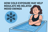
How Cold Exposure May Help Regulate MS-Related Mood Swings
Mood swings are a common but overlooked challenge in multiple sclerosis (MS). This article explores how cold exposure—like cold plunges and showers—may help regulate emotional ups and downs by calming the nervous system, reducing inflammation, and boosting mood-enhancing chemicals. Learn how to use this natural tool safely to support your mental and emotional resilience with MS.
-

MS Fatigue Toolkit: Why Cold Plunges Deserve a Spot in Your Daily Routine
Fatigue is one of the most debilitating symptoms of multiple sclerosis (MS)—often invisible, misunderstood, and overwhelming. While no single tool can eliminate it, building a personalized fatigue management toolkit can make life more manageable. One surprising contender? Cold plunges. In this article, we explore why cold water immersion might be the refresh button your nervous system needs—and how to safely make it part of your MS fatigue routine.
-

Cold Therapy vs. Heat Therapy for MS: Which One Helps More?
Managing multiple sclerosis (MS) often means navigating symptoms like fatigue, spasticity, pain, and nerve dysfunction. But when it comes to using temperature-based therapies, there’s a question many patients face: Should I be using cold or heat? In this in-depth guide, we explore the benefits, risks, and best use cases of cold therapy vs. heat therapy for MS.
-

From the Tub to the Brain: How Cold Plunges Might Support Cognitive Clarity in MS
Cognitive fog is one of the most frustrating symptoms of multiple sclerosis (MS). But could cold plunges—those bracing dips into icy water—offer a surprising path to mental clarity? This article explores the emerging science behind cold exposure, brain function, and how a cold tub might help people with MS sharpen focus, lift brain fog, and reset their nervous system.
-

Why MS Patients Are Turning to Cold Plunges for Relief and Resilience
Cold plunges are no longer just for elite athletes and wellness influencers. A growing number of people with multiple sclerosis (MS) are turning to cold water immersion to ease symptoms, build nervous system resilience, and find calm in the chaos of chronic illness. This article explores why—and how—you might want to give it a try.
-

Cold Plunge Therapy: A Hidden Gem for People with MS?
Cold plunge therapy—once the domain of elite athletes and biohackers—is gaining attention among people with multiple sclerosis (MS). Could it help reduce inflammation, calm the nervous system, and ease MS symptoms like fatigue and spasticity? In this article, we dive deep into the science, benefits, safety, and practical application of cold plunges for MS recovery and symptom relief.
-
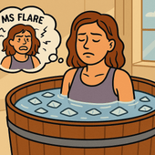
Finding Relief in the Midst of a Flare
MS flares can leave you feeling overwhelmed, exhausted, and mentally foggy. Cold water therapy is emerging as a promising tool to help reset the body and mind after a flare. This article explores how cold exposure supports recovery, calms the nervous system, and can be safely added to your daily routine.
-

How Cold Exposure May Help Regulate MS-Related Mood Swings
Mood swings in multiple sclerosis (MS) can feel like emotional whiplash—one moment calm, the next overwhelmed, angry, or hopeless. While medications and therapy help, many people with MS are exploring natural strategies to support emotional balance. One surprising tool gaining attention? Cold exposure. In this article, we explore how cold plunges and other forms of cold therapy may regulate the nervous system, stabilize mood, and offer emotional relief for people with MS.
-

How to Build an At-Home MS Recovery Corner (with Cold Plunge Setup)
Create your personal MS recovery oasis at home—complete with a cold plunge setup. Learn how to design a space that supports healing, reduces inflammation, and helps you manage symptoms naturally.
-

The Role of Temperature Regulation in MS: Why Cooling Matters


